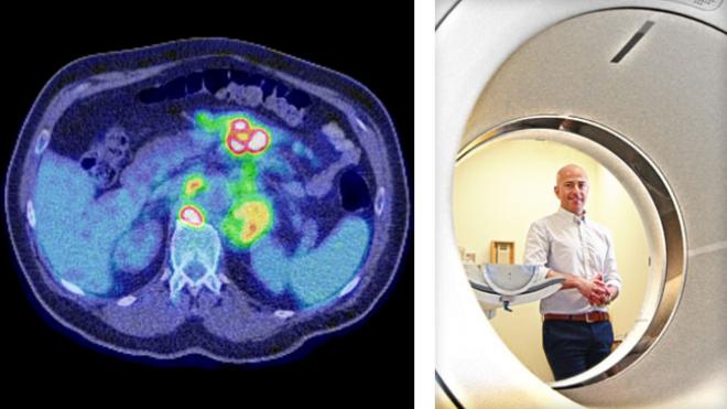
If every picture tells a story, then two pictures have the potential to tell a more complete story.
This appears to be true when two very different types of medical images—computed tomography (CT) and positron-emission tomography (PET) scans—are superimposed to help diagnose cancer.
While a CT scan provides information about the location of and structure of a tumour, a PET scan can visualize fast-growing cancerous cells. Combined, the two images can create a more accurate snapshot of the location and severity of a patient’s cancer—characteristics that are used by physicians to determine the ‘stage’ of a cancer.
Dr. Ur Metser recently led a study that provided the most conclusive evidence to date that PET and CT are a powerful combination for staging lymphoma—a common type of blood cancer. The study was performed in collaboration with Cancer Care Ontario and cancer centres throughout southern Ontario.
He compared the effect of using combined PET/CT to that of CT alone at diagnosis on the management and outcomes of 850 Ontarians with lymphoma.
The researchers found that combined PET/CT enabled the detection of advanced disease in 18% more patients than CT alone and changed treatment plans for 40% of patients. Importantly, PET/CT was linked to improved survival in patients with aggressive non-Hodgkin lymphoma.
“In this study, we show that combined PET/CT enables doctors to more accurately stage lymphomas at diagnosis and thus design effective treatment plans,” says Dr. Metser. “Our findings also suggest that the use of combined PET/CT to inform disease management for certain lymphomas can lead to improved patient survival.”
IMPROVING CLINICAL CARE THROUGH RESEARCH
While the use of combined PET/CT for staging lymphomas was recommended by an international committee of experts in 2013, little prospective evidence existed to support the need for the extra PET scan at the time.

Metser U, et al. Radiology. 2019 Feb;290(2):488-495. Supported by the Ministry of Health and Long-Term Care, Cancer Care Ontario and The Princess Margaret Cancer Foundation.


