Home page Description:
Researchers develop a device to help pre-screen women for breast cancer.
Posted On: August 19, 2016

Image Caption:
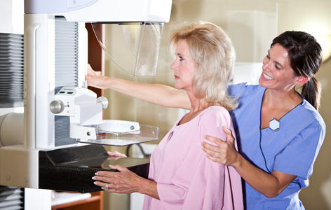
Early detection of breast cancer can be achieved with mammography (pictured); this enables treatments to be started earlier, possibly before the cancer becomes difficult to treat.
Breast cancer is the most common cancer affecting women worldwide. Most screening programs to detect the disease involve mammography, a technique that uses X-ray radiation to visualize breast tissue; however, the value of frequent mammography is under dispute. This is largely due to the tendency to over-screen women, exposing them to potentially unnecessary doses of radiation.
A pre-screening model to predict those that are most likely to benefit from mammography, as well as those that are at risk for breast cancer, would help tailor individual screening plans for women. Knowledge of the density of breast tissue may help to develop such a model because women with dense breast tissue are at increased risk for cancer. Currently, however, breast tissue density is measured with X-ray–based mammography.
To address this issue, PM Senior Scientist Dr. Lothar Lilge and his research team developed a new device to measure breast tissue density without using radiation. The device uses a range of visible and infrared light wavelengths or colours to measure the relative amounts of water, fat and connective tissue in the breast; density is calculated from these data. The researchers found that the device could accurately identify women with high breast tissue density.
“We can now begin to validate this device on a larger population of women,” says Dr. Lilge. “Also, because our device is portable and inexpensive to build, it may be easily incorporated into routine breast cancer pre-screening plans, including those in resource-poor settings.”
This work was supported by the Ontario Ministry of Health and Long-Term Care, the Canadian Institutes of Health Research, the Canadian Breast Cancer Foundation, the University of Toronto and The Princess Margaret Cancer Foundation.
A multi-wavelength, laser-based optical spectroscopy device for breast density and breast cancer risk pre-screening. Walter EJ, Knight JA, Lilge L. Journal of Biophotonics. doi: 10.1002/jbio.201600033. 2016 Jun 7. [Pubmed abstract]


