Home page Description:
Brain imaging ‘fingerprint’ may predict age-related brain damage and intellectual decline.
Posted On: October 10, 2016

Image Caption:
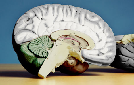
White matter (shown above in white) is located within the central area of the brain; it is involved in learning and facilitates communication between different brain regions.
Magnetic resonance imaging (MRI) provides a detailed view of the soft tissues of the body. When used to visualize the brain, MRI has revealed a phenomenon where high intensity ‘bright signals’ appear in the brain’s white matter. Researchers also call these bright signals leukoaraiosis or white matter hyperintensities (WMHs).
Because they are observed more often in the elderly, WMHs were initially thought to represent structural brain changes that occur as part of normal aging. However, findings have recently linked them to a number of neurological disorders, such as dementia and age-related intellectual decline.
Now, WMHs are the focus of intense study, and it has been proposed that they represent localized ‘mini-strokes’—small brain regions that are damaged as a result of limited blood flow. Despite these hypotheses, much is still unknown about how WMHs form and how to prevent them.
Krembil Senior Scientist Dr. David Mikulis led a study focused on how WMHs form. Using MRI, his team obtained detailed brain images from 45 patients (aged between 50 and 91 years old) at the start of the study and one year into the study.
The researchers found that WMHs formed in brain regions with specific readouts that can be measured with MRI. Specifically, these ‘at risk’ brain regions displayed the following characteristics [MRI readout in brackets]:
This work was supported by the Canadian Stroke Network, the Ontario Research Fund, the Academic Health Science Centre Alternative Funding Plan and the Toronto General & Western Hospital Foundation.
Sam K, Crawley AP, Conklin J, Poublanc J, Sobczyk O, Mandell DM, Venkatraghavan L, Duffin J, Fisher JA, Black SE, Mikulis DJ. Development of White Matter Hyperintensity Is Preceded by Reduced Cerebrovascular Reactivity. Annals of Neurology. doi: 10.1002/ana.24712. 2016 Aug. [Pubmed abstract]
Because they are observed more often in the elderly, WMHs were initially thought to represent structural brain changes that occur as part of normal aging. However, findings have recently linked them to a number of neurological disorders, such as dementia and age-related intellectual decline.
Now, WMHs are the focus of intense study, and it has been proposed that they represent localized ‘mini-strokes’—small brain regions that are damaged as a result of limited blood flow. Despite these hypotheses, much is still unknown about how WMHs form and how to prevent them.
Krembil Senior Scientist Dr. David Mikulis led a study focused on how WMHs form. Using MRI, his team obtained detailed brain images from 45 patients (aged between 50 and 91 years old) at the start of the study and one year into the study.
The researchers found that WMHs formed in brain regions with specific readouts that can be measured with MRI. Specifically, these ‘at risk’ brain regions displayed the following characteristics [MRI readout in brackets]:
- decreased blood flow reserve [cerebrovascular reactivity]
- reduced ‘connectivity’—e.g. reduced ability of the white matter to connect different parts of the brain [fractional anisotropy]
- increases in water distribution [transverse relaxation time]
- increased rates of liquid movement through the tissue, suggestive of tissue injury [mean diffusivity]
This work was supported by the Canadian Stroke Network, the Ontario Research Fund, the Academic Health Science Centre Alternative Funding Plan and the Toronto General & Western Hospital Foundation.
Sam K, Crawley AP, Conklin J, Poublanc J, Sobczyk O, Mandell DM, Venkatraghavan L, Duffin J, Fisher JA, Black SE, Mikulis DJ. Development of White Matter Hyperintensity Is Preceded by Reduced Cerebrovascular Reactivity. Annals of Neurology. doi: 10.1002/ana.24712. 2016 Aug. [Pubmed abstract]

