Home page Description:
The world’s largest multidisciplinary event for photonics meets in San Francisco.
Posted On: March 16, 2017

Image Caption:
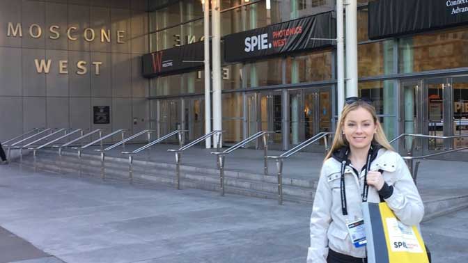
Conference attendee, Sarah Forward, MSc Candidate. Supervisor: Dr. Alex Vitkin, PM.
Conference: SPIE Photonics West (BiOS Exhibition), Jan 28 – Feb 2, 2017, San Francisco, CA, USA.
Conference Highlight: This year’s Photonics West BiOS conference showcased novel optical techniques for biological applications including insights into optogenetics, thick-tissue microscopy, and waveguides for custom light-delivery.
Conference Article:
Photonics West is the biggest photonics conference, bringing together ~20000 students and academics, as well as 1300 photonics companies. There are three main exhibitions to the conference: the BiOS exhibition, which is the largest biomedical optics meeting in the world (where I presented), the LASE exhibition which is focused on cutting-edge optics devices and methods for research applications, and the OPTO exhibition which includes talks on topics such as optoelectronics materials and devices, optical communications and 3-D printing methods.
One of the striking highlights of the conference was presented at the Hot Topics event on thick tissue microscopy or “MUSE” (Microscopy with Ultraviolet Sectioning Excitation). This technique is able to perform histopathology on cut surfaces of thick tissue samples and biopsies using superficially penetrating UV light that requires no need for paraffin embedding, sectioning, or mounting on glass slides. It can be performed on fresh or fixed tissue with less than a minute of prep time and relies solely on common fluorescent stains. The UV light is generated from an LED and penetrates only about 10 microns through tissue, initiating visible-range fluorescent emissions observable through a basic microscope. The images generated are stunningly detailed and colourful and the Levenson group at UC Davis behind this research has even developed software that converts the high-res MUSE images to H&E-style likeness for easy interpretation.
One major theme of the BiOS exhibition was clinical translation. I noticed a lot of the research presented had an emphasis on clinical adoption of new techniques (such as the MUSE work, as well I learned about improving FNA biopsy needles with optics, making routine optical procedures more efficient, calibrating medical endoscopes better, etc.) and so most of the work I saw was very well thought-out from a clinical perspective.


