
Researchers from UHN’s Princess Margaret Cancer Centre have demonstrated how specific cancer cells in a tumour are responsible for driving its growth and spread (also known as cancer propagation).
Despite advances in molecular profiling of cancer cells, disease progression and treatment resistance remain major challenges. This is due in part to the ability of cancer cell clones—groups of cancer cells derived from a single cell—to change their identity and behaviour, a phenomenon known as plasticity.
Scientists have long known that not every cell in a tumour has the ability to regrow the cancer. Only a small number of special cells—called clonogenic cells—can actually start new tumours and keep the cancer going. To cure the disease, these cells must be eliminated. In solid tumours (like breast or lung cancer), finding and studying these key cells has been difficult because they do not have clear markers.
In this study, researchers used a new technique involving genetic barcodes—unique DNA tags that allow scientists to track the descendants of individual cancer cells. Using this method along with advanced single-cell sequencing techniques, they studied over 20,000 individual breast cancer cell clones from 26 patient-derived breast cancer laboratory models.
By tracking these barcoded cells as they divided, the team discovered that fewer than 0.01% of clones were able to regrow tumours. These rare clones divided rapidly and expanded to become the dominant cell type. These clones were also able to reproduce the full gene expression profile of the original cancer model, indicating their ability to drive tumour growth.
In models of basal breast cancer—an aggressive subtype—the tumour-propagating clones showed high plasticity, changing their function and gene activity. The team also identified two distinct cell populations with different clonal growth behaviours and biological features. These results show that propagating clones have the ability to evolve, adapt, and grow into dominant clones that eventually become the majority, all originating from a single barcoded cell.
These findings deepen our understanding of how cancers evolve and resist treatment. This work could lead to therapies that better target these tumour-driving clones, with the approach used here potentially applicable to other solid cancers.
Dr. Long Nguyen, Scientist at Princess Margaret Cancer Centre and Assistant Professor in the Department of Medicine and Medical Biophysics at the University of Toronto, is the first and lead corresponding author of the study.
Dr. Carlos Caldas, Professor of Cancer Medicine at the University of Cambridge, is the co-corresponding author of the study.
This work would not have been possible without the generosity of all the patients who donated samples for the development of breast cancer laboratory models.
This work was supported by The Princess Margaret Cancer Foundation, the Hold’em for Life Early Career Professorship in Cancer Research from the Temerty Faculty of Medicine at the University of Toronto, the European Society For Medical Oncology, Conquer Cancer, The ASCO Foundation, the J.P. Bickell Foundation, the Marathon of Hope Cancer Centres Network, Cancer Research UK, UK Research and Innovation, the National Institute for Health and Care Research, European Research Council, and the Cambridge Commonwealth, European and International Trust.
Dr. Carlos Caldas received research grants (administered by the University of Cambridge) from Genentech, Roche, AstraZeneca, and Servier.
Nguyen LV, Eyal-Lubling Y, Guerrero-Romero D, Kronheim S, Chin SF, Manzano Garcia R, Sammut SJ, Lerda G, Lui AJW, Bardwell HA, Greenwood W, Shin HJ, Masina R, Kania K, Bruna A, Esmaeilishirazifard E, Kolyvas EA, Aparicio S, Rueda OM, Caldas C. Fitness and transcriptional plasticity of human breast cancer single-cell-derived clones. Cell Rep. 2025 May 27;44(5):115699. doi: 10.1016/j.celrep.2025.115699. Epub 2025 May 12.
Patients with complex care needs require more extensive medical, psychological, or social support that often falls outside of standard treatment pathways. A new study from The Institute for Education Research (TIER) at UHN explores how stroke rehabilitation clinicians navigate these challenges and identifies the supports needed to empower adaptive and patient-centred care.
Standardized care pathways are designed to promote efficient, consistent, and evidence-based health care delivery. While effective for many patients, these structured models often lack the flexibility to address the multidimensional and unpredictable needs of individuals with complex conditions, such as those recovering from stroke.
Led by Dr. Alyssa Indar, TIER Educational Investigator, the research team interviewed 22 participants from six accredited Canadian stroke centres. Participants included clinicians from medicine, physical and occupational therapy, nursing, and social work, as well as organizational leaders and health system experts. The study aimed to understand how clinicians deliver care to patients with complex care needs and how this care aligns with organizational expectations and planning.
The findings revealed a disconnect: clinicians reported that most of their patients have complex care needs, while organizational and health system leaders often assume only a small portion require care beyond what is offered in standard pathways. Because leaders influence policy and resource allocation, this disconnect reinforces rigid care models that may not reflect clinical realities. As a result, clinicians frequently develop workarounds to deliver equitable care. However, these strategies are not always sufficient to meet the complex needs of patients and can lead to frustration, moral distress, and burnout among clinicians.
These insights underscore the need for more flexible, supportive health systems that better align policy with patient needs. Empowering care providers to respond effectively to complex care needs can foster more responsive, collaborative, and equitable health care.
Dr. Alyssa Indar, lead author of the study, conducted this work as a PhD student in Dr. Maria Mylopoulos’ ExCEL Lab at the Wilson Centre. Dr. Indar is currently the Director of Scholarship and Innovation in Collaborative Academic Practice and an Educational Investigator at The Institute for Education Research at UHN.
Dr. Maria Mylopoulos, a collaborator on the study, is a Senior Scientist at The Institute for Education Research. She is also a Senior Scientist at The Wilson Centre and an Associate Professor in the Department of Pediatrics and the Institute of Health Policy, Management, and Evaluation at the University of Toronto.
This work was supported by UHN Foundation.
Indar A, Nelson M, Berta W, Mylopoulos M. Exploring perspectives on the management of patients with complex care needs in stroke rehabilitation: An interpretive description study. Health Care Manage Rev. Epub 2025 May 9. doi: 10.1097/HMR.0000000000000440.
For individuals recovering from a stroke or spinal cord injury, restoring hand and arm function is essential to regaining independence. Robot-assisted therapy is an emerging rehabilitation tool that offers consistent and precise movement training. However, its real-world applicability remains limited due to variability in hand use during daily activities outside clinical settings. Researchers from UHN’s KITE Research Institute are addressing this challenge by integrating real-world objects into robot-assisted therapy.
The research team, led by Dr. Milos Popovic, Senior Scientist and Director of the KITE Research Institute, adapted a common clinical assessment tool—the Toronto Rehabilitation Institute-Hand Function Test (TRI-HFT)—for use in robot-assisted therapy. Traditionally, the TRI-HFT involves manipulating everyday objects that require different grips, including a mug, a sheet of paper, a book, a credit card, and a pencil. By redesigning these objects for robotic compatibility, the team enabled the robotic arm to interact with them, allowing the test to be used during therapy.
The team redesigned and 3D-printed 11 TRI-HFT objects. Testing demonstrated that the robotic arm successfully picked up and moved each object, achieving a 100% success rate. Additionally, five participants with normal arm and hand function evaluated the system’s safety and usability, reporting that it was comfortable, engaging, and easy to use.
By combining advanced robotics with traditional rehabilitation tools, this approach enhances the functionality, personalization, and real-world application of robot-assisted therapy. The research explores a new approach to robotic rehabilitation, and the findings lay the groundwork for more effective recovery strategies and improved long-term outcomes. Future research will explore broader clinical applications and adaptability across different robotic systems.
Aisha Raji, a PhD student in Dr. Milos Popovic’s and Dr. Cesar Marquez-Chin's lab, is the first author of the study.
Dr. César Márquez Chin, Scientist at the KITE Research Institute, is a co-author of the study. He is also a Faculty Affiliate at the Institute of Biomedical Engineering at the University of Toronto.
Dr. Milos Popovic, Senior Scientist and Director of the KITE Research Institute, is the senior author of the study. He is also a Professor and Director of the Institute of Biomedical Engineering at the University of Toronto.
This work was supported by UHN Foundation.
Dr. Milos Popovic was a guest editor for the Journal of ‘BioMedical-Engineering OnLine’ for a special issue on the International Conference on Aging, Innovation and Rehabilitation (ICAIR) 2024. To maintain editorial integrity, all standard procedures were strictly followed. The manuscript was handled by independent editors, and Dr. Popovic had no role in the editorial decisions or peer-review.
Raji A, DiNunzio S, Whitmell A, Marquez-Chin C, Popovic MR. Modification of the toronto rehabilitation institute-hand function test for integration into robot-assisted therapy: technical validation and usability. Biomed Eng OnLine. 2025 May 7. doi: 10.1186/s12938-025-01384-7.
Pediatric patients with B-cell acute lymphoblastic leukemia (B-ALL)—a cancer of the blood and bone marrow —can sometimes relapse with features of both B-ALL and a different type of blood cancer, acute myeloid leukemia (AML). In a study published in Nature Cancer, UHN’s Dr. John Dick at Princess Margaret Cancer Centre and Dr. Charles Mullighan at St. Jude Children’s Research Hospital investigated the ability of cells to change from one type to another in B-ALL, and how it affects treatment response.
B-ALL is characterized by the abnormal proliferation of immature lymphoid cells—immune cells that develop into specific types of white blood cells, such as B lymphocytes (B-cells). Categorizing patients into B-ALL subtypes and identifying associated risk levels can predict treatment response and likelihood of relapse. Although pediatric B-ALL cure rates have improved, high-risk children still face poor outcomes, and relapse remains a major cause of death.
Evidence suggests that B-ALL can switch from lymphoid to myeloid lineages after certain immunotherapies or chemotherapy. This phenomenon occurs when a cancerous cell originally classified as lymphoid (e.g., a B-cell precursor) transforms into a myeloid-like cell, such as a granulocyte or macrophage precursor. Malignancies in these lineages lead to different blood cancers, such as ALL from lymphoid cells and AML from myeloid cells. ALL and AML have different molecular features and require different treatment targets.
These findings underscore the importance of understanding B-cell development and lineage switching to predict treatment response. To achieve this, the team analyzed the active and expressed genes (i.e., the transcriptome) of individual leukemia cells from 89 B-ALL patient samples and compared them to normal human B-cell development. To do this, they developed the first comprehensive single-cell reference atlas of normal human B-cell development, spanning over 100,000 cells from various tissue sources.
In constructing this atlas, they discovered that a population of stem cells previously thought to only be capable of producing lymphoid cells (such as B cells) had the hidden ability to produce myeloid cells in the experimental setting.
The researchers found that some B-ALL patient samples contained leukemia cells that highly resemble this population of stem cells with myeloid potential. B-ALL patients who had more of these specific leukemia cells were also more likely to have genomic alterations associated with lineage shifts from lymphoid leukemia to myeloid leukemia at disease relapse.
“Some of these immature lymphoid cells can still develop into myeloid cells,” says Dr. Dick, Senior Scientist at the Princess Margaret Cancer Centre. “This ability, called multipotency, may explain the transition from ALL cases to AML in response to B-cell-specific immunotherapy.”
"We developed a Multipotency Score to describe the abundance of multipotent leukemic cells in patient samples. This score can help predict clinical outcomes,” says Dr. Andy Zeng, co-first author of the study. When tested in independent B-ALL patient datasets, a higher Multipotency Score was associated with higher-risk disease and older age. A high score in pediatric patients was also found to be linked to chemo-resistance and worse overall survival.
“Our research advances our understanding of normal and cancerous B-cell development, which may ultimately enhance risk stratification and therapy development for B-ALL patients,” says Dr. Mullighan, co-corresponding author of the study.
Dr. John Dick, Senior Scientist at Princess Margaret Cancer Centre (PM) and Professor of Molecular Genetics at the University of Toronto (U of T) is a co-senior author of the study. Dr. Dick is also the Helga and Antonio De Gasperis Chair in Blood Cancer Stem Cell Research.
Dr. Charles Mullighan, Co-leader of the Hematological Malignancies Program of the St. Jude Comprehensive Cancer Center is co-senior author of the study.
The co-first authors of the study are Dr. Ilaria Iacobucci, Staff Scientist at St. Jude Children’s Hospital; Dr. Andy Zeng, MD/PhD Candidate at PM and the Temerty Faculty of Medicine at U of T and recent Doctoral Graduate; Dr. Qingsong Gao, Research Scientist at St. Jude Children’s Hospital; and Dr. Laura Garcia-Prat, former Postdoctoral Researcher at UHN and currently a Senior Scientist at Cimeio Therapeutics.
This work was supported by The Princess Margaret Cancer Foundation, St. Jude Children's Research Hospital - ALSAC, the Canadian Institutes of Health Research, National Cancer Institute, Ontario Institute for Cancer Research, International Development Research Centre, Canadian Cancer Society, Terry Fox Research Institute, University of Toronto, University of Toronto’s Medicine by Design, Ontario Government, Alex’s Lemonade Stand Foundation, St. Baldrick's Foundation, Southwest Oncology Group National Clinical Trials Network, and the Henry Schueler 41&9 Foundation.
Ilaria Iacobucci reported consultation honoraria from Arima and travel expenses from Mission Bio and Takara. Charles G. Mullighan received research funding from AbbVie and Pfizer, honoraria from Amgen and Illumina, and royalty payments from Cyrus. He is on an advisory board for Illumina. John E. Dick received research funding from BMS/Celgene and IP licenses from Pfizer/Trillium Therapeutics. For other competing interests, see the manuscript.
Iacobucci I, Zeng AGX, Gao Q, Garcia-Prat L, Baviskar P, Shah S, Murison A, Voisin V, Chan-Seng-Yue M, Cheng C, Qu C, Bailey C, Lear M, Witkowski MT, Zhou X, Zaldivar Peraza A, Gangwani K, Advani AS, Luger SM, Litzow MR, Rowe JM, Paietta EM, Stock W, Dick JE, Mullighan CG. Multipotent lineage potential in B cell acute lymphoblastic leukemia is associated with distinct cellular origins and clinical features. Nat Cancer. 2025 Jun 27. doi: 10.1038/s43018-025-00987-2
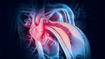
Atherosclerosis—the buildup of cholesterol (called plaques) and inflammation in artery walls—is a leading cause of heart disease and stroke. Researchers at UHN, led by Dr. Kathryn Howe, are studying how the endothelium—the thin layer of cells lining blood vessels—helps drive this process. The team developed the first experimental model to track how endothelial cells (ECs) communicate in organisms and identified EC communication signals that drive carotid artery plaque instability.
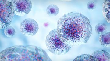
ECs help regulate inflammation and blood flow by releasing tiny particles called extracellular vesicles (EVs). These EVs carry messages in the form of proteins, fats, and genetic material. This dynamic cell-to-cell communication may help drive both plaque development and its sudden complications, such as heart attack or stroke.
However, little is known about these EVs’ biological cargo, their cellular origin or destination, and their functional roles in human plaque formation. In addition, tracking EV transfer from ECs in real time remains challenging. Two studies from Dr. Howe’s team delve into these issues and work to overcome these limitations.
In a study in the journal Circulation Research, the team generated a new preclinical model that uses a glowing green marker to follow EC-derived EVs in real time. This offers a powerful new way to track how EVs move and act within blood vessels—especially in developing plaques—providing a tool for studying EC-driven conditions such as stroke, heart failure, diabetes, cancer metastasis, and aging.
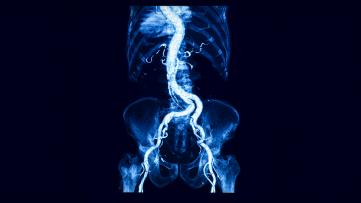
In a second study, published in the journal Arteriosclerosis, Thrombosis, and Vascular Biology, the team analyzed EVs from human carotid artery plaques—where plaque buildup in neck arteries can lead to stroke. In addition to impeding blood flow, the plaques can also become unstable and rupture, releasing material that obstructs an artery and leads to a heart attack or stroke.
The team examined EVs from human carotid plaques, comparing samples from patients with and without symptoms of stroke. Using RNA sequencing and protein analysis, they examined EV cargo and traced their origins and destinations. They found more EVs in plaques than in surrounding tissue—especially in symptomatic patients. These EVs carried different microRNAs and proteins depending on whether plaques were stable or prone to rupture.
In symptomatic plaques, EVs showed more complex communication with nearby cells, particularly endothelial cells, and appeared to promote new blood vessel growth—a process linked to plaque instability.
These findings suggest that EVs help drive disease progression and could be targets for new treatments to prevent stroke and other cardiovascular events. Together, these studies highlight EVs as important drivers of atherosclerosis and introduce a new model to study their role.
For the Circulation Research study:
Mandy Kunze Guo, Doctoral Candidate at UHN and the Institute of Medical Science at the University of Toronto, is the first author of the study.
For the Arteriosclerosis, Thrombosis, and Vascular Biology study:
Dr. Sneha Raju, Medical Resident and recent Doctoral graduate from UHN and the Institute of Medical Science at the University of Toronto, is the first author of the study.
Dr. Kathryn L. Howe, Scientist at the Toronto General Hospital Research Institute (TGHRI) and Associate Professor in the Department of Surgery at the University of Toronto, is the corresponding author for both studies.
Co-author Dr. Jason Fish, Senior Scientist at TGHRI, collaborates extensively with Dr. Howe and together, they co-supervise several graduate students, including those involved in this research. To see more about their laboratories, see https://howeandfishlabs.com.
Dr. Howe holds the Blair Early Career Professorship in Vascular Surgery (University of Toronto) and is supported by the Canadian Institutes of Health Research (CIHR), the Heart and Stroke Foundation of Canada, Wylie Scholar Award from Vascular Cures (2020-2023), the University of Toronto, Peter Munk Cardiac Centre, and the UHN Foundation.
For a more comprehensive list of funding and list of competing interests, see the manuscripts.
Raju S, Turner ME, Cao C, Abdul-Samad M, Punwasi N, Blaser MC, Cahalane RME, Botts SR, Prajapati K, Patel S, Wu R, Gustafson D, Galant NJ, Fiddes L, Chemaly M, Hedin U, Matic L, Seidman MA, Subasri V, Singh SA, Aikawa E, Fish JE, Howe KL. Multiomic Landscape of Extracellular Vesicles in Human Carotid Atherosclerotic Plaque Reveals Endothelial Communication Networks. Arterioscler Thromb Vasc Biol. 2025 May 29. doi: 10.1161/ATVBAHA.124.322324.
Guo MK, Scipione CA, Breda LCD, Prajapati K, Raju S, Botts SR, Abdul-Samad M, Patel S, Yu G, Dudley AC, Fish JE, Howe KL. Tracking Endothelial Extracellular Vesicles in a Mouse Model of Atherosclerosis. Circ Res. 2025 Jun 6;136(12):1629-1631. doi: 10.1161/CIRCRESAHA.124.326024. Epub 2025 Apr 23. PMID: 40265255; PMCID: PMC12136386.
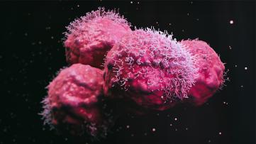
On June 12, 2025, more than 250 in-person attendees—and over 120 virtual participants—gathered at Toronto’s MaRS Discovery District to celebrate the launch of UHN’s newest research collaborative centre: the Collaborative Centre for Immunology to Immunotherapy (Ci2i). The inaugural Ci2i Symposium brought together leading experts in immunology, cell therapy, virology, and immunotherapy for a day of networking, scientific dialogue, and learning.
The event opened with Drs. Tracy McGaha, Chair of the Ci2i Symposium Planning Committee, Brad Wouters, Executive Vice President of Science and Research at UHN, and Pam Ohashi, Chair of Ci2i. Dr. McGaha took the stage first to welcome attendees and set the tone for an exciting day ahead, followed by Drs. Wouters and Ohashi who spoke about the significance of the centre’s launch and shared their vision for the future of immune-related research at UHN.
Throughout the day, researchers from the University of Toronto and UHN presented their work, highlighting the breadth and depth of ongoing research in immunology, virology, immunotherapy, and related fields. Attendees were encouraged to ask questions and engage in thoughtful discussions with the presenters.
Presenters from UHN’s Toronto General Hospital Research Institute included:
● Dr. Brian Coburn, Senior Scientist
● Dr. Slava Epelman, Senior Scientist
● Dr. Stephen Juvet, Scientist
● Dr. Sonya MacParland, Senior Scientist
Presenters from UHN’s Princess Margaret Cancer Centre included:
● Dr. Naoto Hirano, Senior Scientist
● Dr. Tracy McGaha, Senior Scientist
● Dr. Linh Nguyen, Head, Centre for Cell Manufacturing
● Dr. Trevor Pugh, Senior Scientist
● Dr. Ben Wang, Head, Centre for Integrative Immune Analysis
Other presenters a part of TeamUHN included:
● Dr. Gideon Hirschfield, Director, Autoimmune Liver Disease Program, Francis Family Liver Clinic and Toronto Centre for Liver Disease
● Dr. Elmar Jaeckel, Medical Director, Ajmera Transplant Centre
Presenters from the Department of Immunology at the University of Toronto included:
● Dr. Dana Philpott, Associate Chair, Research
● Dr. Alberto Martin, Professor
● Dr. Jennifer Gommerman, Professor
Two international keynote speakers—Dr. Huji Xu, from Shanghai Changzheng Hospital, and Dr. Donna Farber, from Columbia University—bookended the day’s presentations. These internationally recognized researchers provided a global perspective on immune science, showcased their field-defining work, and applauded the Ci2i for its focus on innovation and collaboration.
The symposium also featured a powerful talk by Charlotte Grad, a Ci2i patient partner and former stage 4 non-Hodgkin lymphoma patient treated at the Princess Margaret Cancer Centre. As a CAR-T cell therapy recipient, Charlotte shared her journey, reminding the audience of the real-world impact of immune-based research. “I am here today because of you,” she said to a captivated audience. Her story underscored the life-changing impact that immunology and immunotherapy research at UHN has—and will continue to have with the establishment of the Ci2i.
Ci2i extends its sincere thanks to everyone who helped make the day such a success. This includes the Ci2i Symposium Planning Committee, Firdaus Bakharia, Sara Lamorte, and Ci2i Project Manager Ksenia Meteleva; Dr. Tracy McGaha, Chair of the Ci2i Symposium Planning Committee; Drs. Robert Inman and Naoto Hirano, Co-Chairs of Ci2i; Pam Ohashi, Chair of Ci2i; the Ci2i Executive Committee; and the many volunteers.
The Ci2i team also thanks the Symposium’s five sponsors and three exhibitors:
● Title Presentation Sponsor: 10x Genomics
● Morning Keynote Speaker Sponsor: CDD Vault
● Evening Presentation Sponsors: Bio-Rad Laboratories, and Sanofi
● Patient Partner Sponsor: Cytek Biosciences
● Exhibitors: STEMCELL Technologies, Miltenyi Biotech, and Olink
Abdominal aortic aneurysms (AAAs) are a silent but deadly threat and a leading cause of death in men over 65 in Canada. Currently, there are no treatments to prevent the growth and rupture of these aneurysms. New research from UHN has uncovered how AAAs form and the impact of smoking, potentially opening the door to targeted treatments.
Although initially asymptomatic, AAAs are defined as a swelling of the abdominal aorta—the portion of the aorta that carries blood from the heart to the abdomen. Progressive weakening and dilatation (being stretched or widened) of the vessel wall make it more likely to rupture, which is a surgical emergency and results in death in 50% to 85% of patients. There is limited understanding of how AAAs form and how risk factors such as smoking and arterial plaque buildup contribute to clinical outcomes.
A team led by UHN’s Dr. Clinton Robbins and collaborators used pre-clinical lab models to mimic these risk factors: cigarette smoke exposure and high cholesterol. They found that cigarette smoke drove aneurysm formation by damaging the cells that line the aorta.
Genetic sequencing techniques revealed that this damage triggered inflammation and caused immune cells called macrophages to gather within arterial plaque, where they release enzymes that degrade the elastic fibres of the aortic wall—eventually causing it to expand and rupture like a balloon.
The study also identified a specific type of inflammatory macrophage that was consistent across both pre-clinical lab models and humans, and was central to this destructive process.
“This research sheds light on how smoking worsens AAA and pinpoints the immune cells behind the destruction,” says Dr. Clinton Robbins, Senior Scientist at Toronto General Hospital Research Institute. “It could open the door to new treatments that target these harmful pathways—potentially stopping aneurysms before they burst and become fatal.”
Co-first authors of the study include: Dr. Danya Thayaparan, former Postdoctoral Researcher at McMaster University and current Scientific Advisor at GSK; Dr. Takuo Emoto, Researcher at Kobe University Graduate School of Medicine and former Postdoctoral Researcher at Toronto General Hospital Research Institute (TGHRI); Dr. Aniqa B. Khan, Doctoral Candidate at TGHRI; Dr. Rickvinder Besla, former Doctoral Student at TGHRI and current Senior Scientist at Merck; Dr. Mahmoud El-Maklizi, Postdoctoral Researcher at TGHRI; and Homaira Hamidzada, Doctoral Student at TGHRI.
Drs. Emoto and Khan are also co-corresponding authors on the paper.
Dr. Ken-ichi Hirata, Researcher at Kobe University and Dr. Martin R. Stampfli, Professor at McMaster University, are co-senior authors of the study.
Dr. Clinton S. Robbins, Senior Scientist at Toronto General Hospital Research Institute and Associate Professor in the Department of Laboratory Medicine and Pathobiology at the University of Toronto, is a co-corresponding and co-senior author of the study. Dr. Robbins is a Tier 1 Canada Research Chair in Cardiovascular Immunology and the Peter Munk Chair in Aortic Disease Research at the University Health Network.
This work was supported by the Canadian Institutes of Health Research, the Flight Attendants Medical Research Institute, the Japan Society for the Promotion of Science, the Japan Agency for Medical Research and Development, the MSD Life Science Foundation, the Japan Foundation for Applied Enzymology; and the Naito Foundation, University of Toronto’s Medicine by Design, and the Government of Canada, and UHN Foundation.
Thayaparan D, Emoto T, Khan AB, Besla R, Hamidzada H, El-Maklizi M, Sivasubramaniyam T, Vohra S, Hagerman A, Nejat S, Needham-Robbins CE, Wang T, Lindquist M, Botts SR, Schroer SA, Taniguchi M, Inoue T, Yamanaka K, Cui H, Al-Chami E, Zhang H, Althagafi MG, Michalski A, McGrath JJC, Cass SP, Luong D, Suzuki Y, Li A, Abow A, Heo R, Pacheco S, Chen E, Chiu F, Byrne J, Furuyashiki T, Husain M, Libby P, Okada K, Howe KL, Heximer SP, Yamashita T, Wang B, Rubin BB, Cybulsky MI, Roy J, Williams JW, Crome SQ, Epelman S, Hirata KI, Stampfli MR, Robbins CS. Endothelial dysfunction drives atherosclerotic plaque macrophage-dependent abdominal aortic aneurysm formation. Nat Immunol. 2025 May;26(5):706-721. doi: 10.1038/s41590-025-02132-8.
Research conducted at UHN's research institutes spans the full spectrum of diseases and disciplines, including cancer, cardiovascular sciences, transplantation, neural and sensory sciences, musculoskeletal health, rehabilitation sciences, and community and population health.
Learn more about our institutes by clicking below: